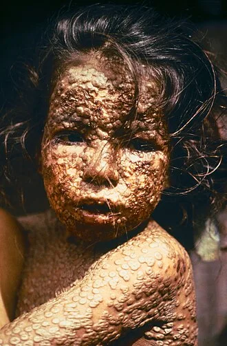
The initial symptoms of the disease included fever and vomiting. This was followed by formation of ulcers in the mouth and a skin rash. Over a number of days, the skin rash turned into the characteristic fluid-filled blisters with a dent in the center. The bumps then scabbed over and fell off, leaving scars. The disease was transmitted from one person to another primarily through prolonged face-to-face contact with an infected person or rarely via contaminated objects.
Prevention was achieved mainly through the smallpox vaccine. Once the disease had developed, certain antiviral medications could have helped, but such medications did not become available until after the disease was eradicated. The risk of death was about 30%, with higher rates among babies. Often, those who survived had extensive scarring of their skin, and some were left blind.
The earliest evidence of the disease dates to around 1500 BCE in Egyptian mummies. The disease historically occurred in outbreaks. It was one of several diseases introduced by the Columbian exchange to the New World, resulting in large swathes of Native Americans dying. In 18th-century Europe, it is estimated that 400,000 people died from the disease per year, and that one-third of all cases of blindness were due to smallpox.
Smallpox is estimated to have killed up to 300 million people in the 20th century and around 500 million people in the last 100 years of its existence. Earlier deaths included multiple European monarchs. As recently as 1967, 15 million cases occurred a year. Inoculation for smallpox appears to have started in China around the 1500s.
Europe adopted this practice from Asia in the first half of the 18th century. In 1796, Edward Jenner introduced the modern smallpox vaccine. In 1967, the World Health Organization intensified efforts to eliminate the disease. Smallpox is one of two infectious diseases to have been eradicated, the other being rinderpest (a disease of even-toed ungulates) in 2011.
The term "smallpox" was first used in England in the 16th century to distinguish the disease from syphilis, which was then known as the "great pox". Other historical names for the disease include pox, speckled monster, and red plague.
Classification
There are two forms of the smallpox. Variola major is the severe and more common form, with a more extensive rash and higher fever. Variola minor is a less common presentation, causing less severe disease, typically discrete smallpox, with historical death rates of 1% or less. Subclinical (asymptomatic) infections with variola virus were noted but were not common.
In addition, a form called variola sine eruptione (smallpox without rash) was seen generally in vaccinated persons. This form was marked by a fever that occurred after the usual incubation period and could be confirmed only by antibody studies or, rarely, by viral culture. In addition, there were two very rare and fulminating types of smallpox, the malignant (flat) and hemorrhagic forms, which were usually fatal.
Ordinary
At least 90% of smallpox cases among unvaccinated persons were of the ordinary type. In this form of the disease, by the second day of the rash the macules had become raised papules. By the third or fourth day, the papules had filled with an opalescent fluid to become vesicles. This fluid became opaque and turbid within 24–48 hours, resulting in pustules.
By the sixth or seventh day, all the skin lesions had become pustules. Between seven and ten days the pustules had matured and reached their maximum size. The pustules were sharply raised, typically round, tense, and firm to the touch. The pustules were deeply embedded in the dermis, giving them the feel of a small bead in the skin.
Fluid slowly leaked from the pustules, and by the end of the second week, the pustules had deflated and began to dry up, forming crusts or scabs. By day 16–20 scabs had formed over all of the lesions, which had started to flake off, leaving depigmented scars.
Ordinary smallpox generally produced a discrete rash, in which the pustules stood out on the skin separately. The distribution of the rash was most dense on the face, denser on the extremities than on the trunk, and denser on the distal parts of the extremities than on the proximal. The palms of the hands and soles of the feet were involved in most cases.
Confluent
Sometimes, the blisters merged into sheets, forming a confluent rash, which began to detach the outer layers of skin from the underlying flesh. Patients with confluent smallpox often remained ill even after scabs had formed over all the lesions. In one case series, the case-fatality rate in confluent smallpox was 62%.
Modified
Referring to the character of the eruption and the rapidity of its development, modified smallpox occurred mostly in previously vaccinated people. It was rare in unvaccinated people, with one case study showing 1–2% of modified cases compared to around 25% in vaccinated people. In this form, the prodromal illness still occurred but may have been less severe than in the ordinary type.
There was usually no fever during the evolution of the rash. The skin lesions tended to be fewer and evolved more quickly, were more superficial, and may not have shown the uniform characteristic of more typical smallpox. Modified smallpox was rarely, if ever, fatal. This form of variola major was more easily confused with chickenpox.
Malignant
In malignant-type smallpox (also called flat smallpox) the lesions remained almost flush with the skin at the time when raised vesicles would have formed in the ordinary type. It is unknown why some people developed this type. Historically, it accounted for 5–10% of cases, and most (72%) were children. Malignant smallpox was accompanied by a severe prodromal phase that lasted 3–4 days, prolonged high fever, and severe symptoms of viremia.
The prodromal symptoms continued even after the onset of the rash. The rash on the mucous membranes (enanthem) was extensive. Skin lesions matured slowly, were typically confluent or semi-confluent, and by the seventh or eighth day, they were flat and appeared to be buried in the skin. Unlike ordinary-type smallpox, the vesicles contained little fluid, were soft and velvety to the touch, and may have contained hemorrhages.
Malignant smallpox was nearly always fatal and death usually occurred between the 8th and 12th day of illness. Often, a day or two before death, the lesions turned ashen gray, which, along with abdominal distension, was a bad prognostic sign. This form is thought to be caused by deficient cell-mediated immunity to smallpox. If the person recovered, the lesions gradually faded and did not form scars or scabs.
Hemorrhagic
Hemorrhagic smallpox is a severe form accompanied by extensive bleeding into the skin, mucous membranes, gastrointestinal tract, and viscera. This form develops in approximately 2% of infections and occurs mostly in adults. Pustules do not typically form in hemorrhagic smallpox. Instead, bleeding occurs under the skin, making it look charred and black, hence this form of the disease is also referred to as variola nigra or "black pox".
Hemorrhagic smallpox has very rarely been caused by variola minor virus. While bleeding may occur in mild cases and not affect outcomes, hemorrhagic smallpox is typically fatal. Vaccination does not appear to provide any immunity to either form of hemorrhagic smallpox and some cases even occurred among people that were revaccinated shortly before. It has two forms.
Early
The early or fulminant form of hemorrhagic smallpox (referred to as purpura variolosa) begins with a prodromal phase characterized by a high fever, severe headache, and abdominal pain. The skin becomes dusky and erythematous, and this is rapidly followed by the development of petechiae and bleeding in the skin, conjunctiva and mucous membranes.
Death often occurs suddenly between the fifth and seventh days of illness, when only a few insignificant skin lesions are present. Some people survive a few days longer, during which time the skin detaches and fluid accumulates under it, rupturing at the slightest injury. People are usually conscious until death or shortly before.
Autopsy reveals petechiae and bleeding in the spleen, kidney, serous membranes, skeletal muscles, pericardium, liver, gonads and bladder. Historically, this condition was frequently misdiagnosed, with the correct diagnosis made only at autopsy. This form is more likely to occur in pregnant women than in the general population (approximately 16% of cases in unvaccinated pregnant women were early hemorrhagic smallpox, versus roughly 1% in nonpregnant women and adult males). The case fatality rate of early hemorrhagic smallpox approaches 100%.
Late
There is also a later form of hemorrhagic smallpox (referred to late hemorrhagic smallpox, or variolosa pustula hemorrhagica). The prodrome is severe and similar to that observed in early hemorrhagic smallpox, and the fever persists throughout the course of the disease. Bleeding appears in the early eruptive period (but later than that seen in purpura variolosa), and the rash is often flat and does not progress beyond the vesicular stage.
Hemorrhages in the mucous membranes appear to occur less often than in the early hemorrhagic form. Sometimes the rash forms pustules which bleed at the base and then undergo the same process as in ordinary smallpox. This form of the disease is characterized by a decrease in all of the elements of the coagulation cascade and an increase in circulating antithrombin.
This form of smallpox occurs anywhere from 3% to 25% of fatal cases, depending on the virulence of the smallpox strain. Most people with the late-stage form die within eight to 10 days of illness. Among the few who recover, the hemorrhagic lesions gradually disappear after a long period of convalescence. The case fatality rate for late hemorrhagic smallpox is around 90–95%.
Pregnant women are slightly more likely to experience this form of the disease, though not as much as early hemorrhagic smallpox.
Signs and symptoms
The initial symptoms were similar to other viral diseases that are still extant, such as influenza and the common cold: fever of at least 38.3 °C (101 °F), muscle pain, malaise, headache and fatigue. As the digestive tract was commonly involved, nausea, vomiting, and backache often occurred. The early prodromal stage usually lasted 2–4 days.
By days 12–15, the first visible lesions – small reddish spots called enanthem – appeared on mucous membranes of the mouth, tongue, palate, and throat, and the temperature fell to near-normal. These lesions rapidly enlarged and ruptured, releasing large amounts of virus into the saliva.
Variola virus tended to attack skin cells, causing the characteristic pimples, or macules, associated with the disease. A rash developed on the skin 24 to 48 hours after lesions on the mucous membranes appeared. Typically the macules first appeared on the forehead, then rapidly spread to the whole face, proximal portions of extremities, the trunk, and lastly to distal portions of extremities.
The process took no more than 24 to 36 hours, after which no new lesions appeared. At this point, variola major disease could take several very different courses, which resulted in four types of smallpox disease based on the Rao classification: ordinary, modified, malignant (or flat), and hemorrhagic smallpox. Historically, ordinary smallpox had an overall fatality rate of about 30%, and the malignant and hemorrhagic forms were usually fatal.
The modified form was almost never fatal. In early hemorrhagic cases, hemorrhages occurred before any skin lesions developed. The incubation period between contraction and the first obvious symptoms of the disease was 7–14 days.
Cause
Smallpox is caused by infection with variola virus, which belongs to the family Poxviridae, subfamily Chordopoxvirinae, genus Orthopoxvirus.
Evolution
The date of the appearance of smallpox is not settled. It most probably evolved from a terrestrial African rodent virus between 68,000 and 16,000 years ago. The wide range of dates is due to the different records used to calibrate the molecular clock. One clade was the variola major strains (the more clinically severe form of smallpox) which spread from Asia between 400 and 1,600 years ago.
A second clade included both alastrim (a phenotypically mild smallpox) described from the American continents and isolates from West Africa which diverged from an ancestral strain between 1,400 and 6,300 years before present. This clade further diverged into two subclades at least 800 years ago.
A second estimate has placed the separation of variola virus from Taterapox (an Orthopoxvirus of some African rodents including gerbils) at 3,000 to 4,000 years ago. This is consistent with archaeological and historical evidence regarding the appearance of smallpox as a human disease which suggests a relatively recent origin.
If the mutation rate is assumed to be similar to that of the herpesviruses, the divergence date of variola virus from Taterapox has been estimated to be 50,000 years ago. A strain that dates from c. 1650 has been shown to be basal to the other presently sequenced strains.
Virology
Variola virus is large and brick-shaped and is approximately 330 nanometers by 260 nm, with a single linear double stranded DNA genome 186 kilobase pairs (kbp) in size and containing a hairpin loop at each end. It infects only humans in nature. Both enveloped and unenveloped virions are infectious. Infection with either the major or minor strain confers immunity against the other.
Variola major, the more common of the two strains, caused the more clinically severe illness and accounted for the great majority of smallpox's historical mortality. The genome of variola major virus is about 186,000 base pairs in length. It is made from linear double stranded DNA and contains the coding sequence for about 200 genes.
The center of the genome contains the majority of the essential viral genes, including for structural proteins, DNA replication, transcription, and mRNA synthesis. The ends of the genome vary more across strains and species of orthopoxviruses. These regions contain proteins that modulate the hosts' immune systems, and are primarily responsible for the variability in virulence across the orthopoxvirus family.
Gene expression occurs entirely within the cytoplasm of the host cell, and follows a distinct progression during infection. About half of the viral genome is transcribed prior to the replication of viral DNA. The first set of expressed genes are transcribed by pre-existing viral machinery packaged within the infecting virion.
These genes encode the factors necessary for viral DNA synthesis and for transcription of the next set of expressed genes. DNA replication in variola virus takes place within the cytoplasm of the infected cell. Recombination of the genome occurs within actively infected cells. The products include transcription factors for transcribing genes for new virions, as well as viral RNA polymerase and other essential enzymes for new viral particles. These proteins are packaged into new infectious virions.
Research
Two live samples of variola major virus remain, one in the United States at the CDC in Atlanta, and one at the Vector Institute in Koltsovo, Russia. Research with the remaining virus samples is tightly controlled, and each research proposal must be approved by the WHO and the World Health Assembly (WHA). Most research on poxviruses is performed using the closely related Vaccinia virus as a model organism.
Vaccinia virus, which is used to vaccinate for smallpox, is also under research as a viral vector for vaccines for unrelated diseases.
The genome of variola major virus was first sequenced in its entirety in the 1990s. The complete coding sequence is publicly available online. The current reference sequence for variola major virus was sequenced from a strain that circulated in India in 1967. In addition, there are sequences for samples of other strains that were collected during the WHO eradication campaign.
A genome browser for a complete database of annotated sequences of variola virus and other poxviruses is publicly available through the Viral Bioinformatics Resource Center.
Genetic engineering
The WHO currently bans genetic engineering of the variola virus. However, in 2004, a committee advisory to the WHO voted in favor of allowing editing of the genome of the two remaining samples of variola major virus to add a marker gene. This gene, called GFP, or green fluorescent protein, would cause live samples of the virus to glow green under fluorescent light.
The insertion of this gene, which would not influence the virulence of the virus, would be the only allowed modification of the genome. The committee stated the proposed modification would aid in research of treatments by making it easier to assess whether a potential treatment was effective in killing viral samples. The recommendation could only take effect if approved by the WHA.
When the WHA discussed the proposal in 2005, it refrained from taking a formal vote on the proposal, stating that it would review individual research proposals one at a time. Addition of the GFP gene to the Vaccinia genome is routinely performed during research on the closely related vaccinia virus.
Controversies
The public availability of the variola virus complete sequence has raised concerns about the possibility of illicit synthesis of infectious virus. Vaccinia, a cousin of the variola virus, was artificially synthesized in 2002 by NIH scientists. They used a previously established method that involved using a recombinant viral genome to create a self-replicating bacterial plasmid that produced viral particles.
In 2016, another group synthesized the horsepox virus using publicly available sequence data for horsepox. The researchers argued that their work would be beneficial to creating a safer and more effective vaccine for smallpox, although an effective vaccine is already available. The horsepox virus had previously seemed to have gone extinct, raising concern about potential revival of variola major and causing other scientists to question their motives.
Critics found it especially concerning that the group was able to recreate viable virus in a short time frame with relatively little cost or effort. Although the WHO bans individual laboratories from synthesizing more than 20% of the genome at a time, and purchases of smallpox genome fragments are monitored and regulated, a group with malicious intentions could compile, from multiple sources, the full synthetic genome necessary to produce viable virus.
Transmission
Smallpox was highly contagious, but generally spread more slowly and less widely than some other viral diseases, perhaps because transmission required close contact and occurred after the onset of the rash. The overall rate of infection was also affected by the short duration of the infectious stage. In temperate areas, the number of smallpox infections was highest during the winter and spring.
In tropical areas, seasonal variation was less evident and the disease was present throughout the year. Age distribution of smallpox infections depended on acquired immunity. Vaccination immunity declined over time and was probably lost within thirty years. Smallpox was not known to be transmitted by insects or animals and there was no asymptomatic carrier state.
Transmission occurred through inhalation of airborne variola virus, usually droplets expressed from the oral, nasal, or pharyngeal mucosa of an infected person. It was transmitted from one person to another primarily through prolonged face-to-face contact with an infected person.
Some infections of laundry workers with smallpox after handling contaminated bedding suggested that smallpox could be spread through direct contact with contaminated objects (fomites), but this was found to be rare. Also rarely, smallpox was spread by virus carried in the air in enclosed settings such as buildings, buses, and trains.
The virus can cross the placenta, but the incidence of congenital smallpox was relatively low. Smallpox was not notably infectious in the prodromal period and viral shedding was usually delayed until the appearance of the rash, which was often accompanied by lesions in the mouth and pharynx. The virus can be transmitted throughout the course of the illness, but this happened most frequently during the first week of the rash when most of the skin lesions were intact.
Infectivity waned in 7 to 10 days when scabs formed over the lesions, but the infected person was contagious until the last smallpox scab fell off.
Concern about possible use of smallpox for biological warfare led in 2002 to Donald K. Milton's detailed review of existing research on its transmission and of then-current recommendations for controlling its spread. He agreed, citing Rao, Fenner and others, that "careful epidemiologic investigation rarely implicated fomites as a source of infection"; noted that "Current recommendations for control of secondary smallpox infections emphasize transmission 'by expelled droplets to close contacts (those within 6–7 feet)'"; but warned that the "emphasis on spread via large droplets may reduce the vigilance with which more difficult airborne precautions [i.e. against finer droplets capable of traveling longer distances and penetrating deeply into the lower respiratory tract] are maintained".
Mechanism
Once inhaled, the variola virus invaded the mucous membranes of the mouth, throat, and respiratory tract. From there, it migrated to regional lymph nodes and began to multiply. In the initial growth phase, the virus seemed to move from cell to cell, but by around the 12th day, widespread lysis of infected cells occurred and the virus could be found in the bloodstream in large numbers, a condition known as viremia. This resulted in the second wave of multiplication in the spleen, bone marrow, and lymph nodes.
Diagnosis
The clinical definition of ordinary smallpox is an illness with acute onset of fever followed by a rash characterized by firm, deep-seated vesicles or pustules in the same stage of development without other apparent cause. Before the availability of laboratory testing, doctors could diagnose smallpox by touch alone, due to the distinctively hard pustules.
Microscopically, poxviruses produce characteristic cytoplasmic inclusion bodies, the most important of which are known as Guarnieri bodies, and are the sites of viral replication. Guarnieri bodies are readily identified in skin biopsies stained with hematoxylin and eosin, and appear as pink blobs. They are found in virtually all poxvirus infections but the absence of Guarnieri bodies could not be used to rule out smallpox.
The diagnosis of an orthopoxvirus infection can also be made rapidly by electron microscopic examination of pustular fluid or scabs. All orthopoxviruses exhibit identical brick-shaped virions by electron microscopy.
Definitive laboratory identification of variola virus involved growing the virus on chorioallantoic membrane (part of a chicken embryo) and examining the resulting pock lesions under defined temperature conditions. Strains were characterized by polymerase chain reaction (PCR) and restriction fragment length polymorphism (RFLP) analysis.
Serologic tests and enzyme linked immunosorbent assays (ELISA), which measured variola virus-specific immunoglobulin and antigen were also developed to assist in the diagnosis of infection.
Chickenpox was commonly confused with smallpox in the immediate post-eradication era. Chickenpox and smallpox could be distinguished by several methods. Unlike smallpox, chickenpox does not usually affect the palms and soles. Additionally, chickenpox pustules are of varying size due to variations in the timing of pustule eruption: smallpox pustules are all very nearly the same size since the viral effect progresses more uniformly.
A variety of laboratory methods were available for detecting chickenpox in the evaluation of suspected smallpox cases.